Understanding the Uterine Tube: Structure, Function, and Health
The uterine tube, also known as the fallopian tube, plays a crucial role in the female reproductive system. Understanding its structure, function, and health is essential for women’s reproductive health and fertility. In this article, we will explore the uterine tube in detail, covering its anatomy, functions, common disorders, and treatment options.
1. What is the Uterine Tube?
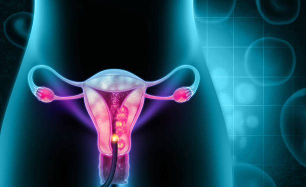
The uterine tube is a pair of slender tubes that connect the ovaries to the uterus. Each woman has two uterine tubes, one on each side of the uterus. These tubes are essential for reproduction, serving as the pathway for eggs to travel from the ovaries to the uterus, where a fertilized egg can implant and develop into a fetus. Understanding the uterine tube’s role is crucial for comprehending how fertility works and the various conditions that can affect it.
2. Anatomy of the Uterine Tube
2.1 Structure
The uterine tube is typically about 10 to 12 centimeters long and is divided into four main parts:
- Fimbriae: These are finger-like projections at the end of the uterine tube nearest to the ovary. The fimbriae help capture the released egg (ovum) during ovulation.
- Infundibulum: This is the funnel-shaped opening of the uterine tube that is closest to the ovary. The infundibulum leads to the ampulla.
- Ampulla: The ampulla is the widest part of the uterine tube and is the most common site for fertilization to occur.
- Isthmus: This is the narrow portion that connects the ampulla to the uterus.
- Uterine Portion: This part of the tube passes through the uterine wall and opens into the uterus.
2.2 Location
The uterine tubes are located in the pelvic cavity. They extend laterally from the uterus towards the ovaries. The tubes are situated above the ovaries and are not directly attached to them; instead, the fimbriae help guide the released egg into the tube.
3. Functions of the Uterine Tube
3.1 Fertilization
The primary function of the uterine tube is to facilitate fertilization. When a sperm cell meets an egg in the ampulla, fertilization occurs. The fertilized egg, now called a zygote, begins to divide and develop as it moves through the uterine tube towards the uterus.
3.2 Transportation of the Ovum
After ovulation, the ovary releases an egg, which is captured by the fimbriae of the uterine tube. The tube’s muscular contractions and cilia (tiny hair-like structures) help move the egg along the tube toward the uterus.
3.3 Support for Early Embryonic Development
The uterine tube provides an environment for the early stages of embryonic development. The fertilized egg remains in the tube for several days, dividing and growing before it reaches the uterus for implantation.
4. Common Disorders of the Uterine Tube
4.1 Ectopic Pregnancy
An ectopic pregnancy occurs when a fertilized egg implants outside the uterus, commonly in the uterine tube. This condition can be life-threatening if not treated promptly, as the growing embryo can cause the tube to rupture, leading to internal bleeding.
Symptoms of Ectopic Pregnancy:
- Abdominal or pelvic pain
- Vaginal bleeding
- Shoulder pain
- Weakness or dizziness
4.2 Pelvic Inflammatory Disease (PID)
PID is an infection of the reproductive organs, often caused by sexually transmitted infections. It can lead to scarring and damage to the uterine tubes, affecting fertility.
Symptoms of PID:
- Lower abdominal pain
- Fever
- Unusual vaginal discharge
- Pain during intercourse
4.3 Tubal Ligation
Tubal ligation, often referred to as “getting your tubes tied,” is a surgical procedure used for permanent contraception. During this procedure, the uterine tubes are cut, tied, or blocked to prevent the egg from reaching the uterus.
5. Diagnosis of Uterine Tube Disorders
5.1 Imaging Tests
Various imaging tests can help diagnose uterine tube disorders. These may include:
- Ultrasound: Uses sound waves to create images of the reproductive organs.
- MRI: Provides detailed images of the body’s soft tissues.
- CT Scan: Can help identify abnormalities in the pelvic area.
5.2 Hysterosalpingography
Hysterosalpingography (HSG) is a specialized X-ray procedure that involves injecting a contrast dye into the uterine cavity. This helps visualize the shape of the uterus and the patency of the uterine tubes.
5.3 Laparoscopy
Laparoscopy is a minimally invasive surgical procedure used to examine the pelvic organs. A small camera is inserted through an incision in the abdomen, allowing the doctor to view the uterine tubes and diagnose any issues.
6. Treatment Options for Uterine Tube Disorders
6.1 Medications
In cases of infections such as PID, antibiotics may be prescribed to eliminate the infection and prevent complications.
6.2 Surgery
Surgical options may be necessary for conditions like ectopic pregnancy or severe damage to the uterine tubes. Surgery may involve removing the ectopic tissue or repairing damaged tubes.
6.3 In Vitro Fertilization (IVF)
For women with blocked or damaged uterine tubes, IVF may be a viable option. In IVF, eggs are retrieved from the ovaries, fertilized in a laboratory, and then implanted directly into the uterus, bypassing the uterine tubes entirely.
7. Maintaining Uterine Tube Health
Maintaining the health of the uterine tubes is essential for reproductive health. Here are some tips for promoting uterine tube health:
- Practice Safe Sex: Using condoms can help prevent sexually transmitted infections, which can lead to PID and other complications.
- Regular Check-Ups: Routine gynecological exams can help identify and address any potential issues early on.
- Healthy Lifestyle: A balanced diet, regular exercise, and avoiding smoking can contribute to overall reproductive health.
8. Conclusion
The uterine tube plays a vital role in female reproduction, facilitating fertilization and the early stages of embryonic development. Understanding its structure, function, and common disorders can empower women to take charge of their reproductive health. By maintaining a healthy lifestyle and seeking regular medical care, women can support their uterine tube health and overall well-being.

