Understanding the Normal ECG Strip: A Comprehensive Guide
Electrocardiography (ECG or EKG) is a crucial tool in modern medicine, allowing healthcare professionals to assess the electrical activity of the heart. A normal ECG strip is a representation of the heart’s rhythm and can provide vital information about a person’s cardiovascular health. This article aims to break down the essential components of a normal ECG strip, interpret its features, and understand its significance in clinical practice.
What is an ECG Strip?
An ECG strip is a graphical representation of the electrical impulses that trigger each heartbeat. It is produced by an electrocardiogram machine, which records the heart’s electrical activity over time. The ECG strip consists of waves, intervals, and segments, each corresponding to specific phases of the heart’s electrical cycle.
The Basics of ECG
- Electrical Activity of the Heart: The heart operates using electrical signals that initiate and coordinate contractions. These signals originate from the sinoatrial (SA) node, commonly referred to as the heart’s natural pacemaker.
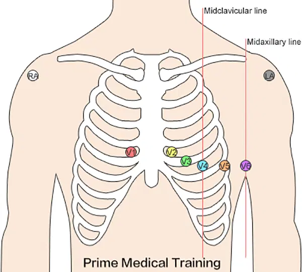
- Recording Process: During an ECG test, electrodes are placed on the skin at specific locations. These electrodes detect the electrical signals produced by the heart, which are then displayed as waves on the ECG strip.
- Waves and Intervals: An ECG strip typically consists of several waves, including:
- P wave: Represents atrial depolarization.
- QRS complex: Represents ventricular depolarization.
- T wave: Represents ventricular repolarization.
Importance of ECG in Clinical Practice
ECGs are essential in diagnosing various heart conditions, including arrhythmias, heart attacks, and other cardiac diseases. A normal ECG strip serves as a reference point, helping healthcare professionals identify abnormalities and make informed decisions regarding patient care.
Components of a Normal ECG Strip
Understanding the normal ECG strip requires familiarity with its key components. The main features include:
1. P Wave
The P wave represents the depolarization of the atria. It is typically a small, rounded wave. The characteristics of a normal P wave include:
- Duration: 0.08 to 0.12 seconds
- Amplitude: Up to 2.5 mm in height
- Shape: Smooth and rounded
2. QRS Complex
The QRS complex indicates ventricular depolarization and is a crucial component of the ECG strip. Its features include:
- Duration: 0.06 to 0.10 seconds
- Amplitude: Can vary but is usually larger than the P wave
- Shape: Typically sharp and pointed
3. T Wave
The T wave represents ventricular repolarization. Its characteristics are:
- Duration: 0.10 to 0.25 seconds
- Amplitude: Generally less than the P wave
- Shape: Asymmetrical and rounded
4. PR Interval
The PR interval is the time taken for electrical impulses to travel from the atria to the ventricles. Its normal duration is:
- Duration: 0.12 to 0.20 seconds
5. QT Interval
The QT interval is the time from the beginning of the QRS complex to the end of the T wave, indicating the duration of ventricular depolarization and repolarization. The normal range is:
- Duration: Varies with heart rate but is generally 0.34 to 0.43 seconds.
6. ST Segment
The ST segment connects the end of the QRS complex to the beginning of the T wave. It is critical in assessing ischemia or injury to the heart muscle. A normal ST segment is:
- Position: On the baseline of the ECG strip.
Interpreting a Normal ECG Strip
Interpreting an ECG strip requires knowledge of normal values and the ability to identify deviations. Here’s a step-by-step approach:
Step 1: Assess the Heart Rate
To calculate the heart rate from the ECG strip, count the number of QRS complexes in a specific time frame. The following methods can be used:
- 6-Second Method: Count the number of QRS complexes in a 6-second strip and multiply by 10.
- Large Box Method: Count the number of large boxes between two QRS complexes and divide 300 by that number.
Step 2: Determine Rhythm Regularity
Check the spacing between the QRS complexes to assess rhythm regularity. A normal rhythm shows consistent spacing, while irregular rhythms indicate potential issues.
Step 3: Measure Intervals
Measure the PR and QT intervals to ensure they fall within the normal ranges. Any deviations may suggest underlying heart problems.
Step 4: Examine Waveforms
Look for the shape and height of the P waves, QRS complexes, and T waves. Ensure they are consistent with normal values.
Step 5: Identify Any Abnormalities
Compare the strip against established norms. Look for signs of:
- Atrial Enlargement: Enlarged P waves can indicate atrial hypertrophy.
- Ventricular Hypertrophy: Enlarged QRS complexes can suggest ventricular hypertrophy.
- Ischemia: Changes in the ST segment may indicate ischemic heart disease.
Common Abnormalities and Their Implications
A normal ECG strip provides a baseline for identifying abnormalities. Here are some common deviations and their implications:
1. Arrhythmias
Arrhythmias are irregular heartbeats that can manifest as:
- Atrial Fibrillation: Characterized by irregularly irregular QRS complexes and absent P waves.
- Ventricular Tachycardia: Displays a rapid succession of wide and bizarre QRS complexes.
2. Myocardial Ischemia
Myocardial ischemia can manifest as ST segment depression or elevation. Key indicators include:
- ST Depression: Often indicates ischemia, especially during exertion.
- ST Elevation: May signify an acute myocardial infarction (heart attack).
3. Electrolyte Imbalances
Electrolyte imbalances can lead to significant changes in the ECG, such as:
- Hyperkalemia: Tall, peaked T waves and widening QRS complexes.
- Hypokalemia: Flattened T waves and the appearance of U waves.
4. Conduction Delays
Conduction delays can occur within the heart’s electrical conduction system, such as:
- First-Degree AV Block: Prolonged PR interval without dropped QRS complexes.
- Bundle Branch Block: Widened QRS complexes with a characteristic shape.
The Role of ECG in Health Monitoring
ECG strips play an essential role in both emergency and routine health monitoring. Understanding the normal ECG strip can help in several ways:
1. Early Detection of Heart Disease
Routine ECGs can detect early signs of heart disease, allowing for timely intervention. Healthcare providers can monitor changes over time to identify potential risks.
2. Postoperative Monitoring
Patients undergoing surgeries, especially cardiac procedures, require careful monitoring through ECG strips. This ensures that any arrhythmias or complications are promptly addressed.
3. Management of Chronic Conditions
Patients with chronic heart conditions often undergo regular ECG assessments to monitor their health status. This helps in adjusting treatment plans accordingly.
4. Screening for Athletes
Athletes may undergo ECG evaluations to screen for any underlying heart conditions that could pose risks during intense physical activity.
Conclusion
A normal ECG strip is a vital tool for assessing heart health. Understanding its components and how to interpret the information it provides is essential for healthcare professionals and patients alike. Regular monitoring through ECG can lead to early detection and management of various cardiac conditions, ultimately improving patient outcomes.
By being knowledgeable about the normal ECG strip, individuals can engage in better health practices and advocate for their cardiovascular health. Continuous education and awareness about heart health are crucial steps towards reducing the prevalence of cardiovascular diseases worldwide.
In summary, whether you are a healthcare professional, a student, or simply someone interested in learning about heart health, grasping the significance of a normal ECG strip can empower you to take charge of your well-being.

